Wednesday, January 19, 2011

CBCT as a Tool in Endodontic Diagnosis

Cone Beam Computed Tomography (CBCT) is a valuable tool in endodontic diagnosis. The following case illustrates how CBCT provides added diagnostic information not available through traditional 2D imaging.

This patient was referred to our office today after a long week of infection and diagnostic dilemmas. Here's the story...

10 days ago with an ear ache.
9 days ago pt reports pain to chewing & closing teeth together.
8 days ago swelling began. Pt went to ER and was given zithromax, ibuprofen & tylenol #3.
7 days ago swelling increased under tongue and into face.
5 days ago, pt returned to ER where they did a CT scan and found nothing. Pt reports numbness in lip. Pt admitted to hospital and given IV clindamycin. MRI done and "something was found in lower left jaw". Pt started 300mg clindamycin.
Today, patient referred from oral surgery for endodontic consult/vitality testing. Here's how he looked.


Radiographs fairly inconclusive. #18, #19, #20, #21 all normal to percussion, probing and thermal testing.

A small crack noted on the distal marginal ridge of #18. Thermal testing once again indicates a vital pulp. Typically, we would expect a necrotic tooth to be the source of the submandibular swelling that this patient has experienced.

Since tooth #18 is responding normally to thermal testing, we decided to take a CBCT to look for more evidence of the source of infection.

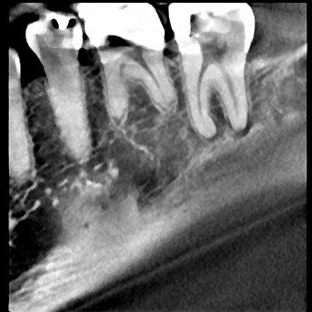
This coronal slice (.25mm) shows radiolucency around the distal root #18. This image is more conclusive than the standard 2D image.

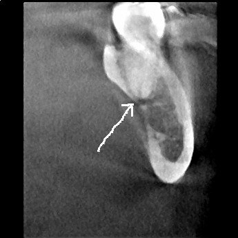
A sagittal slice through the distal root of #18 shows the lesion and its perforation of the lingual plate.

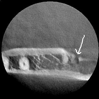
An axial view of the distal root of #18 also shows perforation to the lingual.

These CBCT slices are conclusive enough to revise the pulpal diagnosis to "partially necrotic" and recommend endodontic treatment. It appears that the distal root is necrotic and the infection is spreading through the lingual plate.


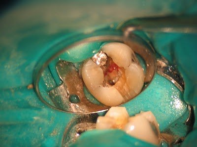
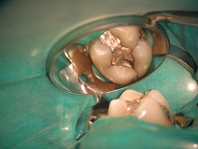
RCT initiated. Upon access, we find vital pulp tissue in the mesial canals, and necrotic pulp tissue in the distal canal.

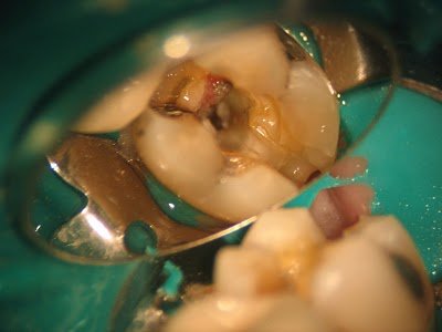
Further removal of the distal crack finds the crack extending down the distal root, below the CEJ. Extraction is recommended.

In endodontic diagnostics, we typically classify pulpal status as:

1. Normal
2. Reversibly Inflammed
3. Irreversibly Inflammed
4. Necrotic

However, things are not always a cut an dry as that. This case illustrates that "partially necrotic" pulp is a possible classification of pulpal status.

Following removal of the tooth, the infection quickly resolved.

CBCT is an important tool for diagnostic imaging in endodontics.

15 comments:

Marcel Caetano said...

Excellent! A true lesson of diagnosis. It shows how much the endodontist must be an artist.

Anonymous said...

I am a dental assistant at Summerlea Dental in Edmonton, AB. I thought this case was very similar to some of the ones that we are presented with. I have to agree with you that CBCT does play an important role in Endodontics. Great Article!

Jon said...

These cases presentations are great. I'm a 3rd year dental student and I can appreciate the art of diagnosis. Keep em coming!

Dr Nikhil Dubey said...

wow.....excellent..

dentist fontana said...

great work..not sure i would have went about it that way though?

dentists in Mississauga said...

its a very indicated and good update on the dentistry business. You can have some of the best results after using some disgnosys of Endodontic.

Jennifer Gibbs said...

Thanks for sharing this interesting case. Pulpal diagnosis can be complex with not all signs and symptoms "adding up" to a clear diagnosis. I think the use of CBCT is also complex and there are costs to the procedures which have to be weighed against the benefits. As there are no guidelines for dentists it is really up to each practitioner how they will use this technology. In this case the CBCT clearly gave us more anatomical information than the PA. However, was that information necessary to get to the diagnosis that would ultimately dictate treatment? I didn't see a mention of the periapical diagnosis in the write up. Usually a cracked tooth with periapical pathology would exhibit pain on biting which could be reproduced with a bite test or percussion. Assuming this were the case, I would feel confident with moving forward with treating #18 given the following evidence:
1. periapical symptoms
2. periapical radiolucency evident on the distal root in PA
3. crack visible on distal ridge
The vital pulp would give me pause but in my mind there would be enough evidence pointing to #18 to move forward with treatment. The diagnosis would be asymptomatic irreversible pulpitis with symptomatic apical periodontitis. Ultimately the CBCT really was of no benefit in this case other than having some great images for documenting an interesting case. Unfortunately the cost to the patient, both financial and the extra exposure to radiation, are not worth the benefit of the scan. This is a great example of why we need good clinical studies so we can know when we really need to go the extra step with expensive imaging.

The Endo Blog said...

Dr. Gibb,

Thank you for your thoughtful comment. I appreciate your point of view.

In the case presented, there was no percussion sensitivity, normal response to cold, normal probings. The diagnosis at that point was: normal pulp & periapex with crack on disto-marginal ridge. The CBCT allowed me to change the pulpal diagnosis to "partially necrotic" and begin treatment without a clinical "guess".

How many times a day do you find yourself telling a patient, "We'll have to open it up and see..." whether that's resorption, decay, cracks, iatrogenic damage, perio defects, sinus involvement etc. I have found that CBCT gives us more information with which to make treatment decisions.

There is additional cost for this improved imaging, but we have kept that reasonable for our patients.

The additional radiation exposure is an important point, however the J. Morita Veraviewepocs 3De is the lowest radiation exposure on the market. It is the equivalant of about 4 periapical films. That is the equivalant of about 4 days worth of normal environmental background radiation exposure.

In this particular case, this patient had already had CT scan (which is a much larger radiation exposure), MRI and been admitted to the hospital. The cost and radiation associated with this lack of appropriate diagnosis cost him much more than the treatment recieved in our office.

I have been asked several times why CBCT would be used in endodontics.
I am finding that a small field of view, low radiation CBCT (such as J. Morita Veraviewepocs 3De) has many applications.

As you can tell, I am very excited about this new technology and am confident it will become as important of a tool as the microscope is in endodontics.

In our practice we continue to use our best judgement to apply this technology as needed for the benefit of our patients.

Thank you again for your comments. I hope you will help us continue these discussions.

Dental Assistant Training said...

hey....it was a very interesting case...i think it will be a great experience for you also...i like the post ..keep it up...

J Chambers said...

Great post! Thanks!

David said...

Excellent technique! Thank you for sharing the information.
Sydney dentist

Justin said...

As an endodontist, I appreciate your thorough and thoughtful approach to this treatment. I take the same approach to fractured teeth and would have also recommended extraction as the most prudent choice.

The CBCT is an interesting tool but one that I have not yet adapted to using. My treatment would have been the same as yours based on almost as convincing RL on the D on the PA image. Strangely, the CBCT looks like it shows a RL on the mesial root as well. Seeing the lesion perforate the cortical plate is definitely cool though.

Between shift shots and traditional film radiographs, I can really get an excellent idea of tooth anatomy and periapical pathology. The CBCT adds something for sure, I am just not sure how much. The case that the salesman always show me are fractured roots (that are obviously fractured on PA) and this case again demonstrates that most cracks are not picked up on CBCT. The other one they always show is the missed ML on maxillary molars or DB on lowar molars (or mid mesial for that matter). I don't need a CBCT to tell me that an molars have 4+ canals; I end up retreating them day in day out, many missed by endodontists more experienced than I. The microscope, some patience, and a cup of six files gets the job done.

I will continue to consider it as an addition to my practice and will check back for more cases. Thank you for your efforts.

I post cases on my facebook page: Alpharetta Endodontics and invite feedback.

-Justin Parente, DMD

The Endo Blog said...

Justin,

Thanks for your comments.

We had that discussion in our practice for about a year before we purchased our CBCT.

We have had a successful endodontic practice for years - before we ever got a CBCT.

I see the CBCT as an additional tool to help us with our endodontic therapy. Being able to see the tooth radiographically from any angle is a valuable image.

I was hoping that by incorporating CBCT into my practice, I could avoid the cases where I tell the patient, "I think this is the problem, we'll have to open it up and see..."

I have not eliminated that, but have become very convinced that CBCT will become as valuable of a tool as the microscope is.

Come to our next seminar in April!

Doylestown Teeth Whitening said...

Excellent blog, witty and informative keep up the good posting!

dentist beverly hills said...

It is just worth it to inspect these situations thoroughly before deciding to do something. I love the way they used the CBCT. Very efficient.