When endodontically treating a tooth experiencing symptoms to percussion, that is symptomatic apical periodontitis or acute apical abscess, it is reasonable and normal to expect those symptoms to persist a few days following treatment. I make it a point to explain to patients that this sensitivity is emanating from the periodontal ligament surrounding the tooth, and, just like a sports injury, ligaments require time to heal following removal of the insult (bacteria in teeth). However, occasionally we evaluate a tooth that has had symptoms to percussion for an extended period of time. In this post, I will discuss the sources of these symptoms and how to diagnose and treat them. I will also show a few cases of persistent apical periodontitis and how they were managed.
Before we get into diagnosis and cases, it is essential to understand the sources of apical periodontitis. With few exceptions, the source of symptoms is either trauma, occlusal or otherwise, or bacteria, apical or otherwise. It really is that simple. It is up to the clinician, through history and exam, to determine the source of symptoms.
Briefly, a common scenario would be a
tooth with an apical radiolucency, sensitivity to percussion, and a lack of cold response. Our diagnosis is necrotic/symptomatic apical periodontitis and the source of both is bacteria. By removing the insulting bacteria with root canal therapy, we can resolve the apical periodontitis and the radiolucency will heal with time. The below images are from a patient with that exact scenario and illustrate an important point.
This #18 clearly has a challenging s-curvature in the apical third of the mesial root. If we are unable to instrument and clean around that curve, we run the risk of bacteria persisting, and consequently, symptoms persisting.
Fortunately, with a little luck, I was able to clean around the curve and the patient’s symptoms resolved within a day of initiation of treatment and calcium hydroxide placement. Also, note the mid-mesial canal. There is an important point here:
With a necrotic tooth, if symptoms persist following treatment, especially if the treatment is deficient in cleaning or rife with errors such as a perforation, then we must suspect bacteria as the source and look to treat the tooth endodontically (possible retreat and/or surgery).
Here is another scenario, this patient was referred for evaluation and treatment of symptoms in the lower left. Symptoms were described as a short, sharp sensitivity to chewing over the past year and a half, and a moderate sensitivity to hot and cold that lingers for a few minutes over the past few months. The patient points to #19 for the chewing sensitivity and farther back for #18. Clearly, we have two issues at hand as an endodontically treated tooth will never cause the type of sensitivity described by the patient.
The previous root canal treatment was done by an endodontist a year and a half ago. This endodontist evaluated the patient and placed him on penicillin and methylprednisone. I must stop here and say that I do not support treating this situation with these medications. I have never felt compelled to prescribe steriods and rarely prescribe antibiotics. It is easy to understand why when we realize that neither of these medications will address the possible sources of the patient’s symptoms. Permanently resolving the inflammation will not happen without removal of the trauma source and systemic antibiotics will not have any effect on bacteria within a root end. Symptoms can be masked, but even a placebo can help a patient’s symptoms. In this case, following medication, there was some improvement in symptoms but they soon returned.
Let us ignore the cold sensitivity for a minute, which was traced to #18, found to linger and be associated with a cracked tooth, and was treated appropriately. What questions are important to ask the patient at this point? Maybe we can deduce the diagnosis of #19 prior to treatment. As it turns out, with more specific questions, I learned that the tooth was cold sensitive prior to root canal therapy, and that these symptoms resolved. I also learned the tooth was actually asymptomatic until the crown was placed a few weeks following treatment. Now, I can proceed to the clinical and radiographic exam knowing that the most likely cause of apical periodontitis is a traumatic occlusion, since even a poor root canal on a vital tooth will likely be asymptomatic for many years before bacteria can negotiate to the apex.
Exam revealed #19 to be sensitive to percussion and bite forces (in MI and with a bite stick), and #18 to be hypersensitive with a prolonged cold response and a fracture. I also located a 5mm ML probing depth with associated erythematous and edematous gingiva. The crown on #19 was bulbous, overhanging the lingual, and flat on the occlusal with wide contacts.
In the below radiographs, we see a short obturation on the mesial and distal roots of #19 with some slight and debatable ligament widening. Not a perfect root canal but unlikely to be the source of symptoms.
Local scaling removed the excess cement and crown recontouring with a diamond bur combined with an occlusal adjustment was performed. The patient returned for treatment of #18 a few days later and all his symptoms from 19 had resolved. A reevaluation in a few months will determine if our treatment worked permanently. Incidentally, here is the completed treatment on #18 (and check out the distal root of 17 curving at the x-ray plane).
The point to be learned from this case is that an accurate and complete history will illuminate possible sources of the problem, exam can confirm that source, and conservative local treatment will often be enough to resolve symptoms. Occlusal trauma is the most common cause of post-treatment apical periodontitis.
Here is a case I treated that exhibited persistent symptoms and is similar in many respects to the one above. The original symptoms and exam were indicative of irreversible pulpitis and extensive recurrent caries was located underneath the buccal margin. I painted a grim picture and recommended extraction. The restorative prognosis was compromised, decay reached into the furcation and alveolar crest. Nevertheless, the referring dentist was confident and capable, and the patient was well informed knowing the risks and that periodontal therapy was in her future. She was anxious to avoid implant treatment due to a prior bad experience with one in another site.
At the second visit of treatment, the patient reported her symptoms to cold had quickly resolved. However, her tooth that was not previously symptomatic to percussion was now hypersensitive to occlusal forces. I had not cleaned long at the first visit, and I had not yet placed the obturation material (not that these usually cause any problems). Likewise, I had merely placed CaOH within the top of the orifices as a barrier to reinfection and not deep into the canals (again, not that this usually causes a problem). The tooth was out of occlusion as well so the only possible source of ligamental inflammation was the buccal area where decay had been removed to the level of the alveolar crest and into the furcation. This area was not well restored yet, merely patched with IRM, and was a definite irritant to the ligament. I cautioned patience, completed treatment, and recommended returning to her general dentist for evaluation and restoration.
When the patient returned for reevaluation a month later, complaining of chewing sensitivity in the temporary crown, I was impressed with the quality of the buccal composite restoration and the buccal gingiva was minimally inflamed. The distal gingiva was inflamed with bleeding on probing. The patient had plans for an evaluation with a periodontist and I again counseled patience with symptoms until all periodontal treatment was complete. The occlusion was found to be heavy in non-working lateral excursion and adjustment relieved the patient’s symptoms in the chair.
The important point here is since I had done the initial root canal treatment, I knew that the tooth was vital preoperatively. There is no way I would consider touching this tooth with any endodontic therapy since I know that I cannot improve upon my root canal treatment. The dental history leads me to look for sources of trauma to the ligament, occlusion and biologic width infringement in this case.
One other common cause that I see of persistent apical periodontitis is when a patient wears an occlusal splint or night guard, dentist fabricated or store bought, that is not well adjusted following a new crown on the tooth. Consider how a kids teeth feel after an orthodontic visit and you will understand the source of these symptoms.
This last case was treated by a general dentist 2-3 years ago and has exhibited persistent symptoms that have only seemed to worsen through multiple occlusal adjustments. The dentist reports an instrument separation in the distal root. Her symptoms had only recently become spontaneous and were described as a minor dull ache or pressure. In discussing the situation with the patient, she remembered the tooth being asymptomatic and the crown having been placed years prior to the root canal treatment. She claimed the referring dentist saw signs of a problem on the radiograph and recommended treatment through the crown. Clinical exam revealed no relevant findings and occlusion appeared well adjusted. A distal shift radiograph, the most important radiograph to take on a maxillary molar, displayed an all too common radiographic finding in maxillary molars treated without a microscope, a radiolucency on the mesial root.
The most common cause of this problem and one that we see every week is a missed MB2 (also called MP or ML) canal. This tooth was unusual in that I speculated a missed MB canal outlined below.
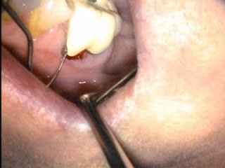
Upon retreatment, the untreated MB canal was found and is seen on the below photograph and radiograph.
Not all cases of persistent apical periodontitis require endodontic retreatment or surgery. In fact, most don’t. By a taking a careful dental history, it is often possible to discern the preoperative diagnosis, which informs how and where bacteria could be causing a problem. It is also possible to create a timeline as to when symptoms first occurred and what events could have initiated them. If symptoms started with restorative work, then carefully examine the occlusion, margins, and gingival health. Only when we have a radiolucency or a smoking gun, like a missed canal, a short obturation in a previously necrotic case, or a case recontaminated with recurrent caries, should we be looking into endodontic therapy as our first choice to solve the problem. Often times, diagnosis is not clear, and I start with the conservative options, occlusal adjustments and periodontal therapy, before proceeding to endodontic therapy. I cannot think of a single instance where I have recommended medication as a primary therapy.
I hope these cases will help those of you out there to think like an endodontist. Some may point out that I have not talked about tooth fractures. I plan to discuss my philosophy on those in future posts. I look forward to any feedback and invite everyone to check out more cases on our facebook page: