Thursday, March 24, 2011

CBCT to Evaluate Apical Lesions

We have been discussing the use of CBCT in the practice of endodontics. There have been questions about whether CBCT is really necessary, or just another cool image. That particular question was one of the biggest we had in our decision to move to CBCT. We also ask that question to ourselves when we recommend a CBCT scan to our patients. However, much like a microscope, until you look through the scope, you often don't know what you are missing. I have found that quite regularly, I will find things that I could not have seen otherwise and it has changed the treatment that I have recommended.

Here's an example of a routine CBCT scan that I did prior to endodontic surgery. This scan gave me added information, that then changed the treatment plan and give us better prognosis.

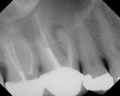
This patient presented for evaluation. The teeth are asymptomatic, but a lesion seen by his general dentist. The lesion is obviously on the MB root of #3, with ledged MB canal. The crown margins looked good and since the MB canal is ledged, we were planning to treat this tooth with an apicoectomy. I recommended a routine, pre-surgical CBCT to evaluate the root anatomy, sinus proximity and buccal bone contours.


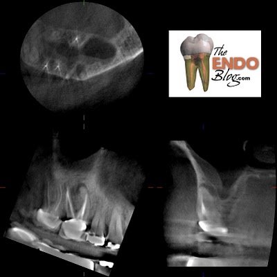
This slice through the MB root shows that there is a missed MB#2 canal.

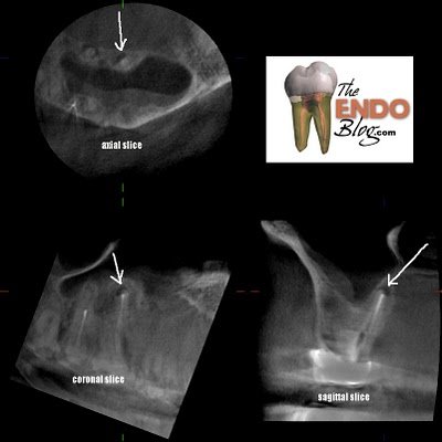
A slice through the palatal root shows a periapical lesion on the palate not visible in the original, pre-op radiograph. This now changes our treatment recommendation. An apicoectomy will resolve the MB issues, but fail to resolve the palatal lesion. This could cause continued problems and lead to the assumption of a failed endodontic surgery, when the palatal root could be the problem.

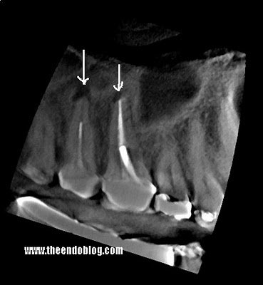
An additional slice through the palatal roots shows that #2 also has a significant periapical lesion requiring treatment.

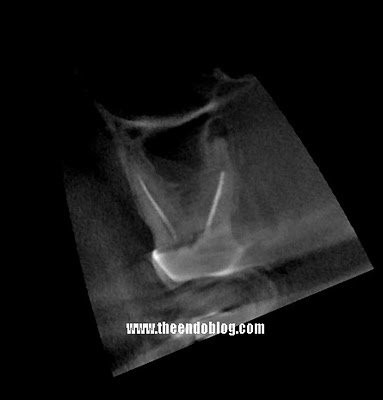
A sagittal view of #2 again shows the extent of the lesion.

While the lesion on the palate of #2 is visible in the original radiograph, there is no doubt about it's presence with the sagittal view above.

In this case the additional information about the palatal lesion on #3 changed the treatment recommendation and will thereby improve the prognosis. Lack of CBCT scan in this case would have led to wrong treatment recommendation.

That being said, I know there are those who will say..."alway retreat first" and you don't need a CBCT scan to make that decision.

CBCT provides improved imaging of the the teeth and periapex. I welcome the added information into the diagnostic and treatment part of my practice. For more information about the application of CBCT into endodontics, the upcoming Inner Space Seminar is right around the corner.

9 comments:

Havre de Grace, MD said...

Good teeth should be everyone's concern. We cannot be caught wearing those ugly smiles. So yes, thank you for this informative post.

Anonymous said...

I'm sure that if you continue to use this advanced imagery you will be able to detect PAR on almost every teeth in this and in almost every mouth. The ethical question is when to treat and is this a true lesion or just apical scarring?Granted there are issues with the quality and esthetics of the original fill but this patient is asymptomatic, re doing No.3 and adding No. 2 into the equation is something to think about. Furthermore if you do look closely at the radiograph presented you can see the palatal PAR on No.2. If we need to find things to do, why don't we look at the existing restorations on this patient, replace them before the endodontic procedure is required.

Justin M. Parente, DMD said...

Beautiful images! I look forward to seeing more. I think that we will likely be adopting CBCT imaging one day in our practice. If we were performing frequent apicoectomies, I would feel more compelled to have the technology.

You already presented my view in your post:

Always retreat first. I have had only 1 indicated apico in the past year and our practice is close to 40% retreatment. We only see cases messed up by GPs.

Never rely on one image to confirm a radiographic diagnosis (especially when surgery is a consideration). Tradigional films with backlight and magnification also help in this respect.

-Justin Parente, DMD
Alpharetta Endodontics

The Endo Blog said...

Anonymous,

Thank you for your thoughts.
In this particular case, these lesions are too big to be scarring.

A proper dental history will insure that teeth are not in the "healing" period following endodontic therapy. If that is the case, and a tooth has a PAR of this size on an asymptomatic tooth, I still think treatment should be recommended. It is a sign of asymptomatic infection.
This technology is not about finding more lesions so we have more teeth to treat, but about finding the things we have not been able to see.

I think it would be a bad idea to do any additional restorative work on #2 with a large, asymptomatic abscess on the palatal root.

If this is my tooth, I would want the endodontic treatment redone, and the PAR healed.

Michael Loftis said...

Everyone needs to listen to what Dr. Hales is reporting on this Blog. I have had the fortunate opportunity to see Dr. Hales operate and evolve his tx philosophy for about 10yrs. I can assure you that he examines all options and would treat as he would do for himself. I have seen Dr. Hales closely research all new technology or technique before adopting anything. As an endodontist myself I can tell you that good endodontist make treatment decisions based on peer reviewed studies. Dr. Hales often cites respected studies to back up his tx choices. This is imperative. One can not only cite anecdotal finding. I think just as important is to consider what you would want for yourself. I think that many DDS settle for one thing for their Pt, but demand something different for themselves. Many would disagee with this statement, but I see it frequently. Many DDS try to tx endo cases that should be refered but when they themselves need tx it must be done by the endodontist. Not the partner they work with everyday. Like I said before we should do for our Pt as we would do for ourselves.


Michael Loftis, DDS

Loftis Endodontics

OdontoCenter said...

Perfect post. I´m Brazil!

Justin said...

Anonymous: It is rarely correct to have restorations ahead of endodontics on a treatment plan. Like Jason mentioned, these asymptoamtic PARLs are clearly not "scars," they are signs of inflammation due to bacterial contamination of the root end and a source of future infection. History can tell us the difference with a high degree of certainty. The term scar, according to the AAE glossary, more accurately describes bone healing after surgery.

apical scar—Dense collagenous connective tissue in the bone at or near the apex of a tooth with a distinctive radiolucent presentation; a form of repair usually associated with a root that has been treated surgically and noted to have perforation of both the facial and lingual osseous cortices.

Justin Parente, DMD
Alpahretta Endodontics

Syneron dental lasers said...

I think that preventive dental care is the key factor in keeping off many dental problems such as Apical Lesions, gum inflammations etc

hellosnackbar said...

After reading this article I'm now of the opinion that any
specialist endodondst needs cbct technology.
I refer my patients for a scan to bone map for implant
placement and wouldn't do it any other way.
What always surprises me is the number of second rate endo treatments that survive for years after.
Clearly this Dr Hales chap is an endo maestro;everything
he has said makes good common sense.